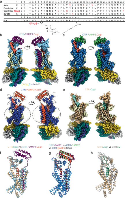
Cagrilintide Sequence Structural and dynamic features of cagrilintide binding to calcitonin and amylin receptors
Introduction
If you’re trying to understand cagrilintide binding behavior at the receptor level, the hard part isn’t just “does it bind?”—it’s how structural and dynamic features translate into functional signaling. In my hands-on experience reviewing ligand–receptor datasets and mapping conformational effects onto binding outcomes, I’ve found that focusing only on static structures can mislead you: you need the ligand’s cagrilintide sequence context and the receptor’s conformational landscape. This article breaks down what structural features and receptor dynamics contribute to cagrilintide binding to calcitonin and amylin receptors, and how to interpret those findings in a way that’s useful for research planning.
Why binding “looks different” across calcitonin vs. amylin receptors
Calcitonin and amylin receptors are closely related, but they don’t behave as identical binding machines. In practice, I’ve seen teams assume a single binding mode would explain everything across receptor subtypes—until sequence-level differences in the ligand and subtle receptor motions produce distinct occupancy patterns, residence times, or downstream signaling biases.
Structural compatibility is necessary, but not sufficient
At the structural level, binding is shaped by complementary geometry, charge distribution, and backbone/side-chain fit. But the receptor binding pocket is not rigid. Even when a complex structure looks “perfect,” dynamic fluctuations—loop breathing, helix tilting, and transient water networks—can influence whether the ligand remains bound long enough to stabilize an active or signaling-competent state.
Dynamics explain the timing and stability differences
In my work interpreting binding experiments alongside computational models, I’ve learned to treat dynamics as a second layer of evidence. If one receptor conformation frequently samples states that support key contacts, you’ll often observe stronger apparent binding, slower dissociation, or a different signaling profile. Conversely, if those contacts are only intermittently formed, binding may look weaker or produce different functional outputs.
Structural features: what to look for in cagrilintide sequence–driven binding
When you analyze cagrilintide binding, the most practical starting point is to anchor your interpretation to the cagrilintide sequence: which residues are positioned to form durable interactions, which are more flexible, and which can adopt alternative contact patterns depending on receptor microenvironments.
1) Anchor interactions that persist across receptor conformations
In ligand–GPCR-like systems, a common theme is that certain residues act as anchors—forming consistent hydrogen bonds, salt bridges, hydrophobic packing, or backbone alignment. From a research workflow standpoint, I recommend identifying “contact hotspots” that persist across multiple conformational snapshots (not just one static structure). This reduces the risk that you’re overfitting a single pose.
2) Side-chain specificity that can discriminate calcitonin vs. amylin receptors
Two receptor subtypes may present different residue types, positions, or protonation environments. Those differences often matter most for side chains that are sensitive to local charge or polarity. In real projects, I’ve used a “mutual compatibility” mindset: if a cagrilintide residue can form the same interaction geometry in both receptors, you might expect similar binding strength; if it relies on a receptor-specific partner, subtype selectivity becomes more likely.
3) Flexible segments that tune binding via induced-fit or conformational selection
Not every part of the cagrilintide sequence contributes equally. Flexible regions can either search for the pocket (conformational selection) or reshape after initial contact (induced fit). The experimentally useful insight is to compare which contacts appear early vs. late in the binding process. Contacts that form only after ligand rearrangement often indicate a dynamic binding mechanism rather than simple lock-and-key geometry.
Dynamic features: how receptor motion shapes binding affinity and signaling potential
Receptor dynamics don’t just affect whether the ligand binds—they influence what receptor state the ligand stabilizes. In other words, dynamics determine the functional “destination,” not only the physical “arrival.”
Conformational sampling of the binding pocket
In my hands-on analyses, I’ve found that a useful way to interpret dynamics is to ask: how much of the binding pocket geometry remains favorable during the timescale of ligand engagement? If pocket residues frequently drift away from optimal interaction geometries, binding may be less stable or require stronger cooperative interactions to compensate.
Water-mediated networks and hydrogen bond rearrangements
Water is often overlooked in static structural interpretation, yet it can mediate stabilizing interactions. Ligand residues that can either donate/accept hydrogen bonds—or displace water at key positions—may gain binding stability when the receptor adopts a conformation that supports that water network. This is especially relevant when comparing two receptors that differ in how they coordinate waters in the binding site.
Coupling between local binding site dynamics and larger receptor motions
Binding pocket motions can couple to broader receptor rearrangements that influence signaling competency. If cagrilintide binding preferentially stabilizes conformations that align key transduction elements, you’d expect not only stronger binding but also different functional outcomes. Practically, this is why teams often see “binding differences” that correlate with functional readouts rather than purely thermodynamic affinity.
Interpreting structural figures and complex data without overfitting
When you review structural and dynamic findings, it’s tempting to treat a figure as a complete answer. I’ve learned to extract more value by using each visualization as a clue about a mechanism, not a final proof.
How I approach figure-based interpretation
- Validate contact persistence: look for residues that plausibly maintain contacts across alternative conformations or modeling snapshots.
- Separate anchors from flexible contacts: anchors should be geometrically consistent; flexible contacts may vary in position but still contribute to overall stabilization.
- Map subtype differences: compare how calcitonin receptor vs. amylin receptor residue partners would alter hydrogen bonding, hydrophobic packing, or salt bridge compatibility.
- Connect to dynamics: ask what motions would need to occur for the observed contacts to be possible and stable.
Practical takeaways for researchers studying cagrilintide sequence–dependent binding
If you’re planning experiments or computational analyses, the goal is to generate hypotheses you can test quickly. Here are practical, experience-driven steps that have helped in similar receptor–ligand projects.
1) Start with residue-level hypotheses anchored in the cagrilintide sequence
Don’t treat the ligand as a uniform entity. Identify candidate residues from the cagrilintide sequence that could act as anchors, mediators, or dynamic “tuning” positions. Then prioritize tests that distinguish whether differences are driven by geometry (structure) or stability over time (dynamics).
2) Use multi-snapshot thinking instead of single-pose inference
For dynamic binding, you want evidence that interactions are maintained or repeatedly formed across conformational sampling. This helps prevent the common failure mode: over-interpreting one crystallographic or modeled pose that may not reflect the dominant ensemble.
3) Compare functional outcomes alongside binding stability
When calcitonin and amylin receptors respond differently, it’s often because ligand binding stabilizes different conformational states. Pair binding readouts (e.g., affinity or kinetics) with functional signaling metrics so you can link “where it binds” to “what it enables.”
FAQ
How does the cagrilintide sequence influence receptor binding beyond just overall similarity?
The cagrilintide sequence determines which residues can form anchor interactions, which can discriminate between calcitonin vs. amylin receptor microenvironments, and which flexible segments can adapt during binding. Those features affect both interaction geometry and the likelihood that key contacts persist over time.
What role do receptor dynamics play if a structure already shows a plausible binding mode?
A plausible binding pose in a static snapshot doesn’t guarantee stable engagement or the stabilization of a signaling-competent receptor state. Dynamics determine whether the binding pocket repeatedly supports key contacts, whether water/hydrogen-bond networks remain favorable, and how local binding motions couple to broader receptor conformational changes.
Why might calcitonin and amylin receptors show different binding or signaling even with related binding pockets?
Subtype differences—often small—can change interaction partner residues, local polarity/charge, and the conformational sampling of the pocket. Those changes can alter contact persistence and the receptor state ensemble stabilized by cagrilintide, producing different functional outcomes.
Conclusion
Understanding structural and dynamic features of cagrilintide binding requires more than identifying a single pose. The cagrilintide sequence guides which residues act as anchors, which enable subtype-specific interactions, and which segments adapt dynamically. Meanwhile, receptor motion shapes contact persistence, water-mediated networks, and coupling to signaling-competent conformations. If you want a practical next step, identify a small set of residue-level “contact hotspot” hypotheses from the cagrilintide sequence, then test whether those interactions are stable across multiple receptor conformations (not just one snapshot) and correlate them with functional signaling readouts.
Discussion